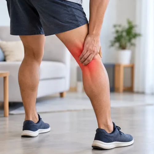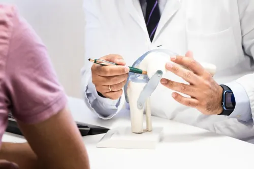
A knee injury is not treated from an MRI label alone: the structure involved, degree of damage, joint stability, and your functional goals all change the plan. By the end, you will know which findings call for urgent assessment, how clinicians choose imaging and treatment, and what progress to track before returning to normal activity.
Key takeaways
- Seek urgent assessment for deformity, inability to bear weight, or a locked knee.
- Do not force a locked knee straight or walk on it after major trauma.
- MRI shows soft-tissue damage; X-ray and CT assess bones and alignment.
- Combined ligament, cartilage, or fracture injuries need a coordinated treatment plan.
Start with triage: which knee injuries need urgent assessment?
A knee injury emergency needs immediate assessment when the joint looks deformed, you have inability to bear weight after significant trauma, or the knee is locked and cannot fully straighten. Do not try to force it straight or walk on it.
| Situation | What to do | Why it matters |
|---|---|---|
| Knee deformity or suspected dislocation | Go to an emergency department now | Bones or the kneecap may be displaced, and a knee dislocation can injure the popliteal artery even when foot pulses initially feel normal |
| New numbness, pale or cold foot, or absent or weaker pulses | Seek emergency care immediately | Nerve or blood-vessel damage can threaten the lower leg |
| Hot, markedly swollen knee with severe pain and very limited movement | Obtain same-day urgent assessment, especially with fever or feeling unwell | Septic arthritis requires joint-fluid testing, antibiotics, and drainage; waiting for an MRI can delay treatment |
| Inability to bear weight, major swelling soon after impact, or inability to walk four steps | Arrange urgent assessment and likely X-rays | These findings raise concern for fracture or a serious internal injury |
| Mild pain with improving swelling, normal shape, and comfortable weight-bearing | Book a routine appointment and limit aggravating activity | A stable sprain can wait if no warning sign develops |
An ACL tear alone is usually assessed urgently but treated as an elective problem after swelling and motion are addressed, not as an emergency. Worsening pain, swelling, numbness, fever, or new deformity changes that plan.
How the injury is identified: examination, X-ray, MRI, and CT
A normal X-ray does not rule out an ACL tear, meniscus injury, cartilage damage, or another internal derangement. Knee injury diagnosis combines the injury mechanism, symptoms, examination findings, and imaging before treatment is chosen.
1. The clinician records how the injury happened: twisting, a direct blow, forced hyperextension, or a fall. They note pain location, immediate swelling, bruising, locking, giving-way, weight-bearing ability, previous knee problems, age, and activity demands.
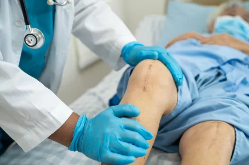
2. The examination compares both knees. It includes swelling, warmth, tenderness, deformity, range of motion, strength, joint-line pain, ligament stability, kneecap tracking, and pulses or sensation. A hot, very swollen knee with severe motion restriction and fever requires urgent joint-fluid aspiration for cell count, Gram stain, and culture to check for septic arthritis.
3. The Ottawa Knee Rule identifies many acute injuries that need radiographs: age 55 or older, fibular-head tenderness, isolated patellar tenderness, inability to bend to 90 degrees, or inability to walk four weight-bearing steps. A knee X-ray is the initial test when fracture is suspected.
4. If radiographs show no fracture but internal damage remains likely, knee MRI without contrast is the preferred next study for ACL or PCL tears, menisci, cartilage, tendons, and bone bruising.
5. CT supplies finer fracture detail when a break is complex, displaced, or being planned for surgery. Clinicians then combine these findings to classify the injury as stable or unstable, partial or complete, and isolated or combined.
How structure and severity change the treatment choice
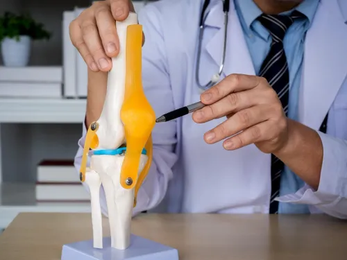
Treatment follows two questions: which structure failed, and whether the knee remains aligned and stable under load. A stable injury can often be protected while pain settles and motion returns; displacement, loss of continuity, or repeated giving-way raises the need for reduction, closer review, or surgery.
- Grade 1 sprain: fibers are stretched, but the ligament remains stable. Use protection, early controlled movement, and strengthening.
- Grade 2 sprain: fibers are partly torn and laxity is present. A hinged brace, protected loading, and rehabilitation come before surgery when the knee remains controllable.
- Grade 3 sprain: the ligament is completely torn or functionally detached. Rehabilitation may suit an isolated injury, but persistent instability, pivoting-sport demands, or combined damage make reconstruction more relevant.
- The partial versus complete tear distinction matters for ACL, PCL, and tendon injuries, but MRI appearance alone does not decide treatment; knee function and examination also guide it.
- A nondisplaced fracture may need immobilization and protected weight-bearing. A displaced fracture, joint-surface step, or unstable alignment may need reduction and fixation.
- Meniscus treatment aims to preserve usable tissue: repair a repairable acute tear, while a displaced fragment causing locking may require limited removal.
- A first-time patellar dislocation often receives reduction, bracing, and rehabilitation. An osteochondral fracture, loose body, or recurrent instability changes the surgical discussion.
- Combined ligament, meniscus, cartilage, or tendon damage can leave the knee unstable even when one injury alone would respond to rehabilitation.
Why isolated and combined injuries follow different plans
A second injury can change the knee’s stability, healing conditions, and safe treatment sequence, so an apparently familiar injury may need a different plan.
An isolated ACL tear in a stable knee might be managed with rehabilitation, while an ACL and meniscus tear may require protection of the meniscus and reconstruction planning, especially if the knee gives way or you want to return to pivoting sport.
An isolated MCL injury often heals with activity modification, controlled motion, strengthening, and a hinged brace. Add an ACL, PCL, or meniscal tear, and the knee can remain unstable; MCL injury treatment then has to address the full combined knee injury rather than the inner ligament alone.
PCL injury treatment also often starts with bracing and rehabilitation when the knee functions stably, but combined ligament damage, persistent instability, or an avulsion can shift the decision toward surgery.
Use these differences as questions at review:
- Is the knee stable during walking and daily tasks, or does it buckle?
- Is there locking from a meniscus tear, cartilage fragment, or loose body?
- Is a fracture displaced or an osteochondral fragment loose after kneecap dislocation?
- Will treating one structure first protect another structure’s healing?
- Do motion, swelling, strength, and balance support rehabilitation, or is instability preventing progress?
A first-time kneecap dislocation often needs reduction, bracing, and rehabilitation, but recurrent instability or a fracture changes the plan. A normal X-ray does not exclude internal damage; MRI can reveal the additional injury driving that change.
How non-surgical treatment is reviewed before the next decision
Non-surgical care is a reasonable first plan when the knee is stable, the injury is suitable for protection and rehabilitation, and you can follow activity limits. Examples include many Grade 1 sprains, selected partial ligament tears, first-time kneecap dislocations without a loose bone fragment, and nondisplaced fractures.
A knee brace and protected weight-bearing may limit stress while knee injury rehabilitation restores movement and control.
Review progress against measurable findings rather than pain alone:
- Swelling should reduce, the knee should straighten fully, and bending should improve without a blocked or locked feeling.
- Walking should become steadier, with less dependence on crutches and less pain during prescribed weight-bearing.
- Strength and single-leg control should improve compared with the uninjured side.
- The knee should not repeatedly give way, buckle, or swell after routine activity.
- Your function should match the next activity level, not just a calendar date.
Continuing non-surgical care makes sense when these markers improve without recurrent instability or a new loss of motion. Persistent locking, repeated giving-way, worsening swelling, inability to progress weight-bearing, or an osteochondral fragment requires reassessment; the plan may shift to further imaging or surgery.
For athletes, return to sport criteria include full motion, controlled swelling, near-symmetrical strength, balance, hopping or landing control, and sport-specific cutting or pivoting drills. Dr. Manohar Rao Ortho can use this type of review to judge whether rehabilitation is succeeding or a different intervention is needed.
Related services
Knee Replacement also known as knee arthroplasty, is a transformative surgical procedure that relieves pain and restores function in severely damaged knee joints. View service → |
Primary Knee Replacement Primary Total Knee Replacement is the most common type of knee surgery performed by Dr. View service → |
Frequently asked questions
Which knee injuries need urgent assessment?
Seek immediate assessment if the knee looks deformed, you cannot bear weight after significant trauma, or the joint is locked and cannot fully straighten. Do not force it straight or walk on it.
How do doctors identify the type and severity of a knee injury?
Doctors combine the injury history with a physical examination. X-rays assess bones, MRI evaluates ligaments, menisci, cartilage, and other soft tissues, while CT provides detailed views of complex fractures and alignment.
Why do combined knee injuries need a different treatment plan?
An isolated injury can target one damaged structure, while combined injuries affect stability, movement, or bone alignment together. Treatment must address the full injury pattern and its effect on knee function.
How is non-surgical knee injury treatment reviewed?
Doctors review pain, swelling, range of motion, strength, stability, weight-bearing ability, and progress with rehabilitation. If the knee remains unstable, locked, painful, or functionally limited, they reassess the diagnosis and next treatment.
Related services
Sports Injury Treatment Sports injuries can significantly hinder an active lifestyle. At our center, Dr. View service → |
meniscal injury treatment Meniscal tears are common knee injuries often resulting from twisting movements or trauma. Dr. View service → |
Cartilage Repair & Regeneration Cartilage Repair & Regeneration focuses on restoring damaged joint cartilage to relieve pain, improve movement, and delay or prevent the progression of arthritis. View service → |